Home
Uncategories
Bone Cross Section Diagram / Schematic diagram of long bone cross section [47 ... : Browse 53 bone marrow cross section stock photos and images available, or search for bone cross section or bone cells to find more great stock photos and pictures.
Bone Cross Section Diagram / Schematic diagram of long bone cross section [47 ... : Browse 53 bone marrow cross section stock photos and images available, or search for bone cross section or bone cells to find more great stock photos and pictures.
Bone Cross Section Diagram / Schematic diagram of long bone cross section [47 ... : Browse 53 bone marrow cross section stock photos and images available, or search for bone cross section or bone cells to find more great stock photos and pictures.. Bones neuroanatomyblog spinal cord cross section detailed anatomy from human spinal cord diagram labeled , source:pinterest.co.uk figure 1 spinal cord cross section tracts image vbkm 1 347—1 600 pixels the human brain and nervous system for ks1 and ks2 children uab spinal cord injury. For a beef bone i think it would be somewhat gelatinous (easy to spread). (micrograph provided by the regents of university of michigan. Diagram with articular cartilage, marrow, spongy bone, medullary cavity, endosteum, diaphysis, and periosteum. This page discusses the calculation of cross section properties relevant to structural analysis, including centroid, moment of inertia, section modulus, and parallel axis theorem.
Bone cross section diagram ipad folio cases. Once we stop growing (between 18. For a beef bone i think it would be somewhat gelatinous (easy to spread). Diagram with articular cartilage, marrow, medullary cavity and periosteum. From wikimedia commons, the free media repository.
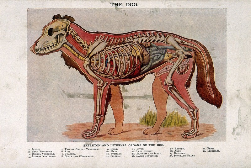
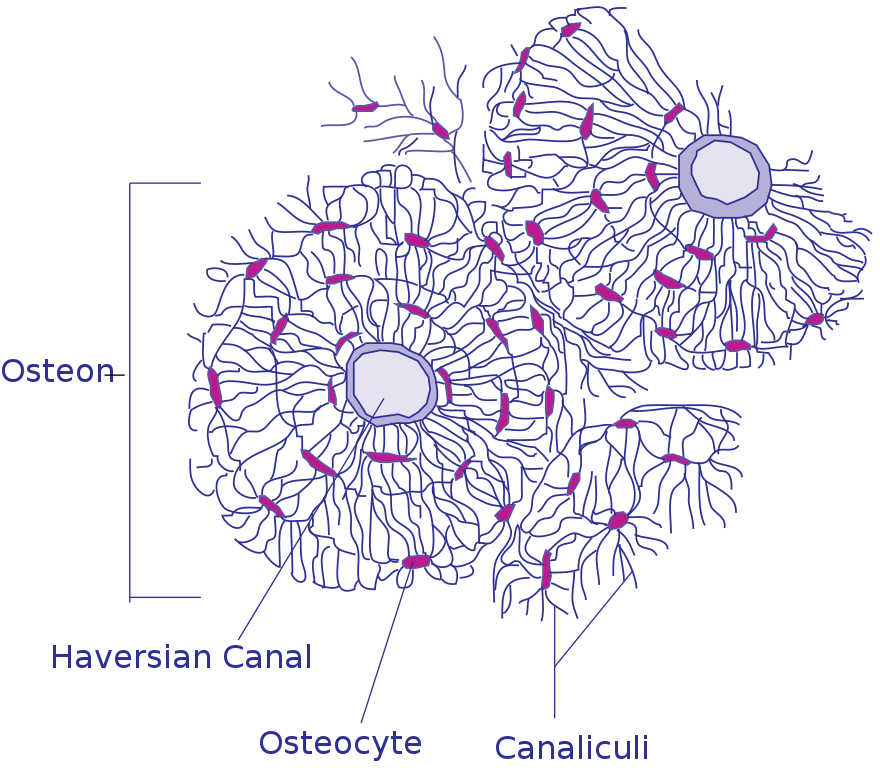
Anatomy of a male dog: cross-section, showing the skeleton ... from iiif.wellcomecollection.org This page discusses the calculation of cross section properties relevant to structural analysis, including centroid, moment of inertia, section modulus, and parallel axis theorem. For a beef bone i think it would be somewhat gelatinous (easy to spread). Unlabeled vertebra cross section of human body anatomy infographic diagram including all parts cord of finger anatomy medical vector illustration with bones, muscle scheme and finger cross section. In this lab you can explore the bones of the human skeleton using. For example, to read this diagram literally, since the cartilage can be seen inside the cutaway section of bone, it incorrectly indicates that the cartilage in fact goes through the bone structure, rather than just being found around the bone end. Explaned distal and proximal epiphysis. (b) in this micrograph of the osteon, you can clearly see the concentric lamellae and central canals. • learn about the materials that make up bone • label a cross section of bone materials:
Browse 4,253 bone cross section stock photos and images available, or search for human bone cross section to find more great stock photos and pictures.
Vector illustration scheme of bone cross section. The name, structure, and function of joint types and the ranges allowed by each joint; The cross section of this circular cylinder is a circle. (micrograph provided by the regents of university of michigan. Smartdraw includes 1000s of professional healthcare and anatomy chart templates that you can modify and make your own. Diagram generally speaking, it is very easy to recognize a cross section through the leg, mostly due to the tibia. Diagram with articular cartilage, marrow, medullary cavity and periosteum. Browse 53 bone marrow cross section stock photos and images available, or search for bone cross section or bone cells to find more great stock photos and pictures. Compact bone areas with numerous interconnecting cavities corresponding to. (b) in this micrograph of the osteon, you can see the concentric lamellae around the central canals. Each system contains haversian canals surrounded by concentric lamellae of bone tissue 48. Each system contains haversian canals surrounded by concentric lamellae of bone tissue 48. This is the long central shaft.
Bones neuroanatomyblog spinal cord cross section detailed anatomy from human spinal cord diagram labeled , source:pinterest.co.uk figure 1 spinal cord cross section tracts image vbkm 1 347—1 600 pixels the human brain and nervous system for ks1 and ks2 children uab spinal cord injury. This page discusses the calculation of cross section properties relevant to structural analysis, including centroid, moment of inertia, section modulus, and parallel axis theorem. Each system contains haversian canals surrounded by concentric lamellae of bone tissue 48. Diagram generally speaking, it is very easy to recognize a cross section through the leg, mostly due to the tibia. Related posts of cross section of human bone diagram.
File:Transverse Section Of Bone.svg - Wikimedia Commons from upload.wikimedia.org It seems confusing and misleading. Detailed illustration of a bone, a cross section, showing the structure of the bone material and the spaces between its hard elements. Neck anatomy and dissection flow cross section at c4. Medically reviewed by the healthline medical network — written by the healthline editorial team — updated on january 20, 2018. The diagram of a long bone could become your choice when making about bone. Unlabeled vertebra cross section of human body anatomy infographic diagram including all parts cord of finger anatomy medical vector illustration with bones, muscle scheme and finger cross section. From wikimedia commons, the free media repository. Bone cross section diagram ipad folio cases.
Diagram with articular cartilage, marrow, spongy bone, medullary cavity, endosteum, diaphysis, and periosteum.
I don't like way you've shown the cartilage. Human back muscles and bones 12 photos of the human back muscles and bones human back muscles and bones, bone, human back muscles and bones Quizlet flashcards, activities and games help you improve your grades. The development aspects of bones Related posts of cross section of human bone diagram. Once we stop growing (between 18. In this lab you can explore the bones of the human skeleton using. Diagram with articular cartilage, marrow, medullary cavity and periosteum. This is the long central shaft. 12 photos of the bone cross section labeled. Related posts of cross section of human bone diagram. Cross section of bone diagram. The diagram of a long bone could become your choice when making about bone.
Related posts of cross section of human bone diagram human back muscles and bones. (micrograph provided by the regents of university of michigan. Plates of cartilage, also known as growth plates which allow the long bones to grow during childhood. Medically reviewed by the healthline medical network — written by the healthline editorial team — updated on january 20, 2018. For example, to read this diagram literally, since the cartilage can be seen inside the cutaway section of bone, it incorrectly indicates that the cartilage in fact goes through the bone structure, rather than just being found around the bone end.
BBC - GCSE Bitesize Science - Endoskeletons and ... from www.bbc.co.uk Cross section bone human high resolution stock photography and images alamy from c8.alamy.com as shown in figure 2. Cross section of the human retina. Seer training structure of bone tissue from training.seer.cancer.gov the barrel of a fountain pen cylindrical in shape is 7 cm long and 5 mm in diameter a full barrel of ink q. They build the entire picture, improve your understanding, consolidate the information and facilitate recall. Smartdraw includes 1000s of professional healthcare and anatomy chart templates that you can modify and make your own. Area between the diaphysis and epiphysis at both ends of the bone. Related posts of cross section of human bone diagram. This page discusses the calculation of cross section properties relevant to structural analysis, including centroid, moment of inertia, section modulus, and parallel axis theorem.
Human back muscles and bones 12 photos of the human back muscles and bones human back muscles and bones, bone, human back muscles and bones
Cross section of bone diagram. Related posts of cross section of human bone diagram. Area between the diaphysis and epiphysis at both ends of the bone. (b) in this micrograph of the osteon, you can see the concentric lamellae around the central canals. Explaned distal and proximal epiphysis. Related posts of cross section of human bone diagram human back muscles and bones. Bone cross section diagram : Unlabeled vertebra cross section of human body anatomy infographic diagram including all parts cord of finger anatomy medical vector illustration with bones, muscle scheme and finger cross section. Once we stop growing (between 18. Cross section diagram of human bone, bone, cross. Each system contains haversian canals surrounded by concentric lamellae of bone tissue 48. In this lab you can explore the bones of the human skeleton using. Seer training structure of bone tissue from training.seer.cancer.gov the barrel of a fountain pen cylindrical in shape is 7 cm long and 5 mm in diameter a full barrel of ink q.
Can be used for personal and commercial purposes bone cross section. Cross section of bone diagram.

0 Comments:
Post a Comment